Hair features
Hair features
Hair identification is most successful when using guard hairs, and is based on recognising and describing the following range of features:
All hairs consist of a basic three layered structure consisting of a central core (medulla) surrounded by a cortex (may be pigmented) and an outer cuticle consisting of overlapping scales (Figure 1). The terminology used in this database to describe hair features is based on that of Brunner and Coman (1974).
The age of the animal, the part of the animal where the hair is growing, the growth stage of a hair and the position along the length of the hair shaft affects the appearance of all five features. (see Figure 2).
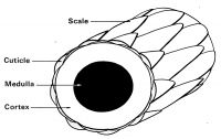
Fig. 1. Magnified representation of basic hair structure
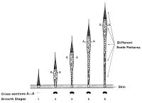
Fig. 2. Five stages in the development of a primary guard hair in the brown rat (Rattus norvegicus)


Types of hair
The coat of a mammal, referred to as a “pelage”, has five different types of hairs (see Figure 3 and Figure 4). A hair sample may have a mixture of hair types present. Record all hair types in the sample. These may be identified as follows:
- Guard hairs – the most important hairs for species identification. These are the larger or coarser hairs forming the main pelage. They tend to be uniform diameter along their length, with a taper toward the tip. Can vary considerably in size (length and diameter) over the pelage, with finer guard hairs indistinguishable from under hairs.
- Overhairs – hairs that are noticeably longer than the main pelage, tend to be circular in cross-section and sparsely scattered over the pelage.
- Underhairs - not useful for species identification. Usually wavy, and shorter and finer than guard hairs. Usually uniform diameter along their length, with a taper toward the tip. Can vary considerably in size (length and diameter) over the pelage.
- Bristle hairs – a course type of hair present in the pelage of some animals such as pigs. The hairs tend to be stout, rigid and uniform diameter along their length.
- Vibrissae – sensory hairs e.g. whiskers.
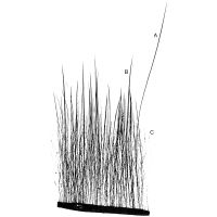
Fig. 3. A tuft of hair from the brown rat (Rattus norvegicus), showing an overhair (A), guard hairs (b) and underhairs (C)
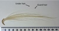
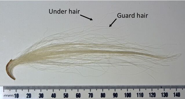
Fig 4: Kurī hair sample showing guard and underhairs.

Hair profile
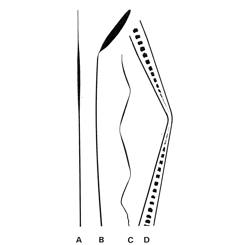
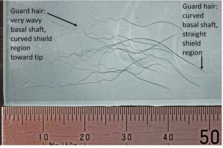
The hair profile is a macroscopic description of the hair as seen by the naked eye or using low magnification (e.g. a magnifying glass. Some profile features are easiest to see when viewed in silhouette (e.g. Figure 5 and Figure 6). Features to note include:
- The maximum length and diameter of the hair/s
- The general shape of the hair/s – e.g. variation in diameter (e.g. Figure 5 A), taper, presence of a shield region (e.g. Figure 5 B)
- Undulations – e.g. straight (e.g. Figure 5 A), simple curve, wavy (e.g. Figure 5 C), very wavy
- Constrictions – note if present or absent
- Colour – note any variations or banding along the length of the hair
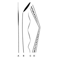
Fig. 5: Some examples of hair profiles. A and B are guard hairs, C is an underhair with constrictions, D represents a magnified view of a constriction in an under hair
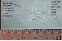
Fig. 6. Brushtail possum (Trichosurus vulpecula) hair profiles. Note shield region seen as thickening of hair diameter toward tip.
Scales
Scales can only be seen using a microscope capable of at least 200x magnification. Three aspects of the scales should be noted and described based on the illustrations in Figure 7:
- The form of the scale margin
- The distance between the scale margins
- The scale pattern
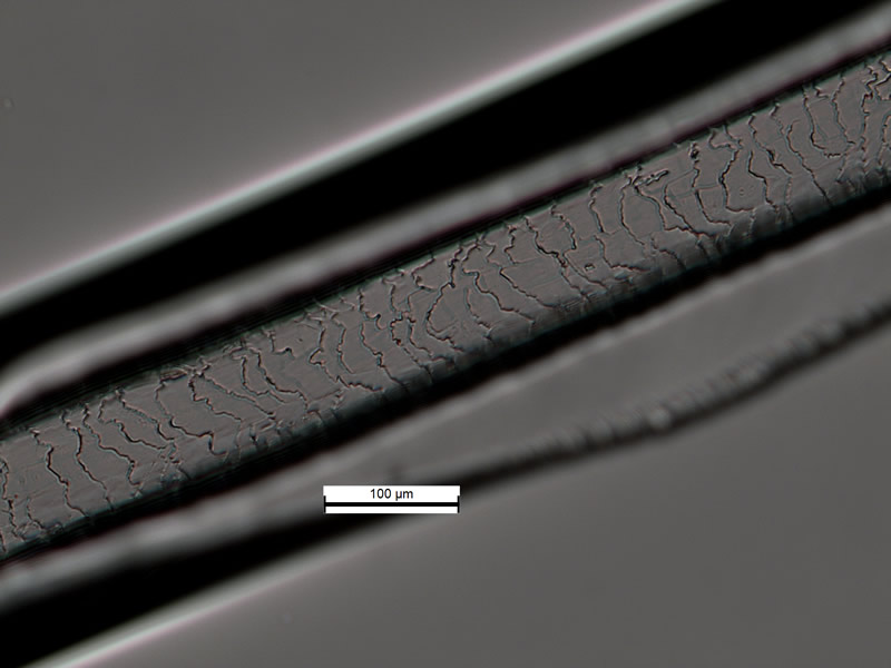
Scales are often observed by taking scale casts from the hair. See Brunner and Coman (1974) for a description of the method used to prepare scale casts. Figure 8 shows a typical scale cast prepared using nail polish. Scales can still be seen without taking a scale cast, by carefully adjusting the focus, contrast, image brightness and illumination mode (transmitted or reflected) of the microscope. See the example in Figure 9.
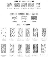
Fig. 7. Various shapes and arrangements of cuticular scales
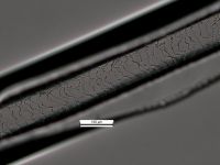
Fig. 8. Scale cast taken from a dog (Canis familiaris) hair.
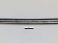
Fig. 9. Scales on dog (Canis familiaris) hair made visible by adjusting microscope settings.

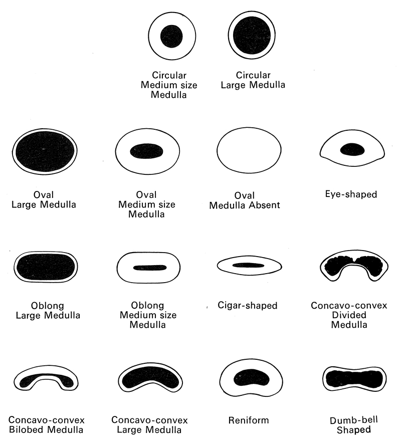
Medulla
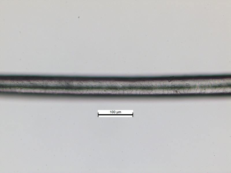
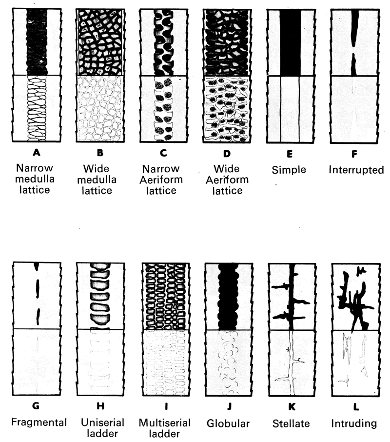
The medulla can only be seen using a microscope capable of at least 200x magnification. A polarising microscope can further enhance the appearance of the medulla. Figure 10 illustrates the aspects of the medulla that should be noted and described; Figure 11 shows a narrow medulla lattice in a dog hair using cross-polarised light on a polarising microscope.

Fig. 10. Medulla types and terminology
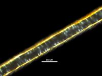
Fig. 11. Narrow medulla lattice in a dog (Canis familiaris) hair, clearly visible using cross polarised light microscopy.

Cross-sections
Hair cross-sections are considered an important diagnostic feature, but may not always be available. Cross-sections should be taken from the widest portion of a well-developed primary guard hair from an adult, to be reliably diagnostic. The technique for preparing cross-sections requires practise. Instructions can be found in Brunner and Coman (1974) or Brunner and Triggs (2002). Figure 12 shows common cross-sectional shapes of hairs. Figure 13 shows the typical appearance of a hair cross-section prepared using the technique described by Brunner and Coman (1974).
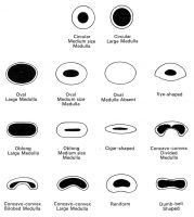
Fig. 12. Some shapes of cross-sections at the widest part of the primary guard hair
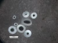
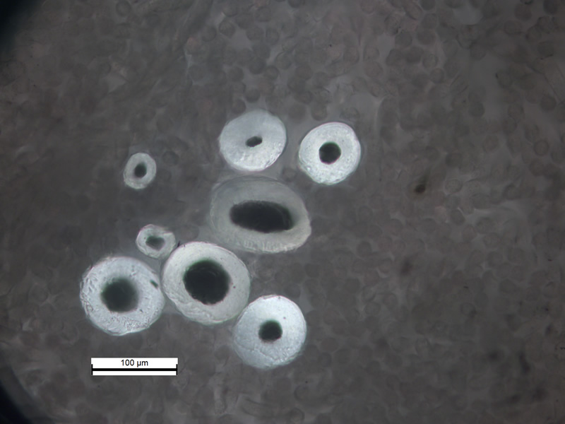
Fig. 13. Cross-section of dog (Canis familiaris) hairs.
Root and tip
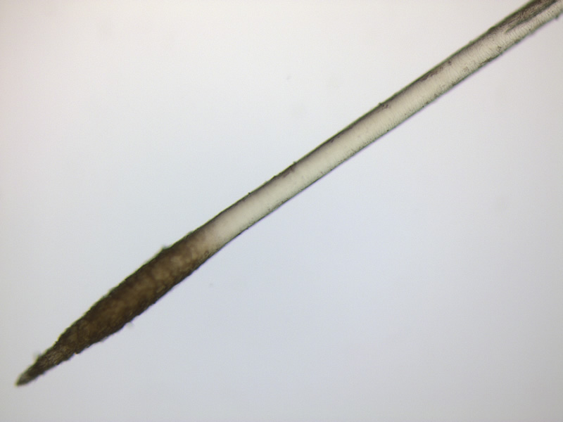
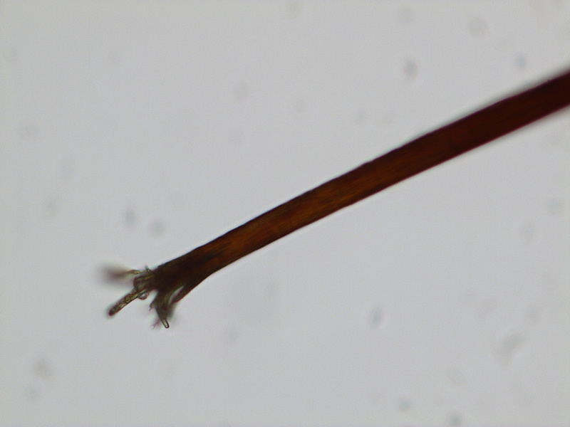
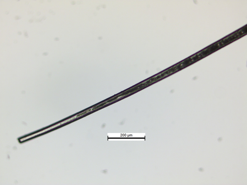
The root of the hair is the enlarged end of the hair embedded in the skin of the animal (Figure 14). In the past, the shape of the root of a hair was thought to be a useful diagnostic feature for identifying some species of animal. However, root shape is not widely accepted as a diagnostic feature, and therefore has not been included in this online resource. Similarly, the tip of a hair can be diagnostic for a limited number of species (e.g. pig (Sus scrofa) hairs can have a frayed tip (Figure 15). When observing the tip of a hair, take care that a damaged hair end (e.g. Figure 16), as is often observed in hairs form artefacts, is not mistaken for flagged hair tip. The true tip of a hair can usually be recognised by the way the medulla appears to taper off close to the tip (Figure 17).
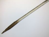
Fig. 14. Typical appearance of the root of a dog hair when viewed with a microscope
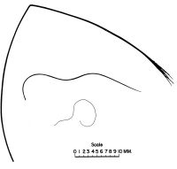
Fig. 15. Pig (Sus scrofa) hair profiles showing the flagged or frayed tip
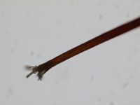
Fig. 16. Typical example of the frayed appearance of a damaged hair endtip

Fig. 17. Typical example of an un-damaged hair tip. Note the medulla becoming narrow and tapering off toward the end of the hair